The Telethon Kids Institute is equipped with a diverse range of histology and microscopy equipment and analysis software to facilitate state-of-the-art imaging and study of intracellular organelles, cells and tissue.
Bright Blue Cancer Analysis Suite![]()
Generously supported by donations from Bright Blue, the Institute provides a suite of equipment encompassing all aspects of tissue processing for histology and pathology and enabling a wide range of microscopy applications including transmitted light (brightfield, phase contrast, differential interference contrast (DIC) and fluorescence-based techniques such as epifluorescence (with spectral unmixing capabilities) and confocal.
We also have capabilities to allow live cell time-lapse microscopy for short-(hours) or long- (days) term experiments. Together, this equipment enables our researchers to visualise cells or tissue to study cell function and how they are affected by changing environmental, physiological and disease conditions such as cancer.
The facility incorporates a range of manual and automated histology and pathology instruments including:
- Leica Automated Vacuum Tissue processor ASP200S
- Leica Tissue paraffin embedder EG1150
- Leica CM1800 cryostat
- Leica RM2135 microtome
- KD3358 Semi-automatic microtome + floatation workstation
- Leica Autostainer XL
Imaging Equipment
-
Nikon C2+ Confocal Microscope
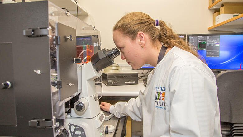 Confocal microscopy is an imaging technique which increases optical resolution and contrast by the use of a small pinhole which collects light only from the plane of focus, eliminating ‘out of focus’ glare and enabling the collection of serial optical sections from thick specimens. Telethon Kids Institute houses a Nikon C2+ system enabling multimode (three colour confocal, transmitted light DIC, epifluorescence imaging), multipoint and time lapse imaging.
Features
- Inverted Nikon Ti-E Microscope.
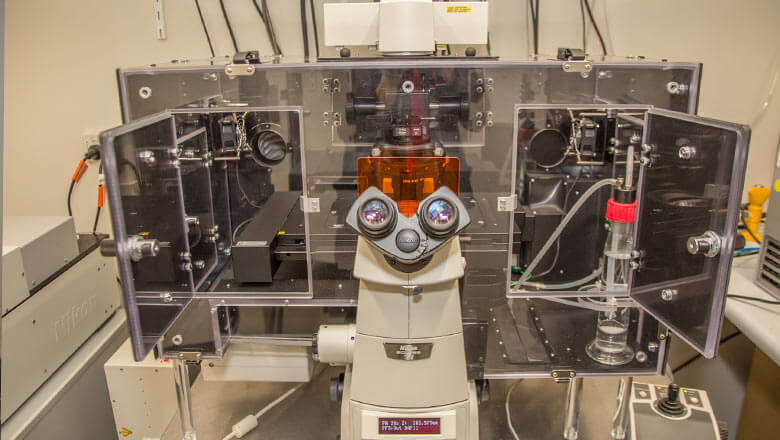 Four solid state lasers-405nm (violet), 488nm (blue), 56nm (yellow) and 638nm (red).
- Three PMT Detectors.
- Nikon Objectives 2x-60x, air, water and oil immersion.
- NIS Elements software controlling all microscope component and enabling image processing and analysis.
- Perfect focus system for live imaging.
- OkoLab live cell imaging chamber.
- Epifluorescent filter cubes suited for imaging a number of fluorophores including DAPI, GFP, FITC, CFP, YFP, TRITC, mCherry.
-
3D Histech Pannoramic MIDI Slide Scanner
-
Nuance Multispectral Imaging System
-
Mantra Multispectral Imaging System
-
Incucyte S3 Live Cell Analysis System
-
Additional Microscopy Systems
Contact us
Please direct general enquiries to our reception on (08) 6319 1000.
For further information about our laboratory equipment, please contact:
Tamara Abel
Microscopy and Equipment Specialist
Phone: (08) 6319 1440
Email: Tamara Abel